

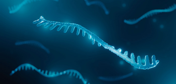
In biology’s standard tale of two acids — deoxyribonucleic (DNA) and ribonucleic (RNA) — RNA is merely the messenger.
Its better-known cousin DNA plays the lead role, issuing genetic instructions for the construction of complex molecules called proteins that do the cells’ work. Recent research, however, shows that specialized proteins that bind to RNA also regulate the messages RNA delivers.
Mutations in the nearly 2,100 genes that encode RNA-associated proteins occur in cancer, neurodegeneration, developmental disorders and other diseases. But it’s been difficult to pin down what each of those proteins is doing within the cell.
Researchers at Fred Hutch Cancer Center invented a new screening method called ReLiC — described recently in the journal Nature Methods — that solves that problem using CRISPR-Cas9, the Nobel-prize winning gene-editing technology.
Instead of knocking out the genes that encode RNA-associated proteins one at a time, ReLiC tests them all at once on a batch of cells, each cell edited so that it’s missing only one key gene.
ReLiC measures how those gene deletions affect various RNA processes that are traditionally difficult to measure at scale.
“This approach has opened up a new way for us to systematically interrogate RNA regulation,” said Arvind (Rasi) Subramaniam, PhD, a researcher in the Basic Sciences Division who joined Fred Hutch 10 years ago and studies how cells make proteins from RNA.
Many things influence RNA, from transcription to translation to degradation
Messenger RNA is a go-between molecule that copies DNA codes for building proteins — a process called transcription — and then delivers the transcripts to the cell’s many protein factories, which are called ribosomes.
In the ribosomes, the RNA sequences are translated into amino acid sequences, which are then used to make proteins.
RNA-associated proteins influence RNA’s function from the moment a messenger RNA molecule is created during transcription to when it is translated in the ribosome all the way to its eventual degradation when it’s no longer needed.
“But which of these proteins in the cell are actually doing the job, and what exactly are they doing?” Subramaniam asked.
One way to find out is to delete a gene that codes for an RNA-associated protein and see what changes.
“You can do it for a few genes but doing it for more than that gets really unwieldy,” he said.
What the field needed was a screening technique flexible enough to identify the functions of thousands of RNA-associated proteins at once without breaking the bank.
As part of his doctoral thesis research, Patrick J. Nugent, PhD, a former student in the Subramaniam Lab, took on that challenge and helped develop the method they call ReLiC (the capital letters stand for RNA-Linked-CRISPR).
Using CRISPR to reveal hard-to-reach RNA processes
The ReLiC method relies on a molecular tool called site-specific integration to deliver a customized set of DNA sequences called a library to a specific location in the human genome.
“The site-specific integration method helps us keep everything the same except the gene that we are deleting,” Subramaniam said. “That’s what makes it powerful. We can knock out about 2,000 human genes in a single Petri dish.”
The library includes CRISPR guide molecules that deliver a molecular scissor called Cas9 to the genes that researchers want to knock out. When the cell repairs the break, it’s usually not good enough to restore the gene’s function, which knocks it out.
ReLiC combines the CRISPR guides with short random sequences called barcodes inserted into RNAs that help the researchers monitor each deletion.
The addition of barcodes helps them keep track of what happens to various RNA processes that are not typically accessible to CRISPR when a gene is knocked out and its relevant proteins aren’t made.
A few days after the Cas9 snippers have done their work, the batch of cells undergoes sequencing, which counts the barcodes in the RNA.
If a barcode becomes more or less abundant, it suggests that the knocked out gene plays an important role in either making the RNA process more or less efficient.
The power of looking at many things at once
The researchers used ReLiC to figure out what proteins were important for translating messenger RNA transcripts into proteins, which occurs in the cells’ ribosomes.
As expected, hundreds of genes encoding ribosomes or the proteins that help start the translation process were important because turning them off with CRISPR impaired how well RNA was translated. Surprisingly, translation was impaired when they knocked out other genes involved in how proteins fold into 3-D shapes or how proteins are destroyed.
The team also discovered a gene that regulates how cells respond to a drug used to treat chronic myeloid leukemia that targets the ribosome to inhibit protein synthesis. While other ReLiC experiments revealed several genes and pathways, this experiment isolated a single gene that made a difference in how effectively the drug works to inhibit protein synthesis.
Using ReLiC, Subramaniam and his colleagues also found hard-to-reach networks of molecular pathways, protein complexes and individual proteins regulating events before and after translation.
These included splicing, which trims nonessential information copied during transcription, and how messenger RNA molecules break down or decay after translation when their work is no longer needed.
Though much can be learned by knocking out one gene at a time, the ability to delete all the relevant genes systematically gives the lab a bird’s eye view of how RNA regulates protein production.
“We can really now ask these big questions about how different RNA processes talk to each other or to the rest of the cell," Subramaniam said. “It has changed the trajectory of my lab’s research. That’s the power of looking at many things at once.”
Co-authors include Fred Hutch researchers Stanley Lee, PhD, in the Translational Science and Therapeutics Division and Andrew Hsieh, MD, in the Human Biology Division.
This research was supported by grants from the National Institutes of Health and the Genomics and Flow Cytometry Shared Resources of the Fred Hutch/University of Washington/Seattle Children’s Cancer Consortium and Fred Hutch Scientific Computing.
This article was originally published August 5, 2025, by Fred Hutch News Service. It is republished with permission.




Comments
Comments