


This post picks up where The Smudged Thumbprint left off. We met with the radiation oncology team to interpret the latest MRI and plan the next steps. As always, I’m writing in the space between medical data and lived experience — trying to make sense of what we see, and what it means to keep living inside it.
A few days ago, I shared about my latest MRI, what the radiologist called a BT-RADS 3a, meaning “likely progression, but possibly treatment effect:
Whit grabbed a pic of a scan image from a recent MRI, an image identified specifically in the radiology report as an image suggestive of the finding: BT RADS 3a, meaning, 3: “imaging worsening (indeterminate/likely progression): worsening imaging findings,” and a: “worsening due to treatment effects.”
What continued in that post was a pragmatic read of circumstances, like the functional decline in my health—something really seems to be going wrong in my body; the image itself, as I described it, a smudged thumbprint, and the statistical likelihood of this aggressive cancer that after nine years, chemo in 2017, 2024, and 2025, radiation in 2016 and 2025, and the three different periods of confirmed growth since 2023, a scan that looked like the one Whitney and I were seeing from last month (September 2025), just seemed to fit the pattern of disease progression—and not treatment effect, as the radiology report suggested.
Why Bring This Up Again?
Like I began the linked post, “This post is for anyone, but it’s probably best suited for other brain tumor families,” I write for the world, but what Glioblastology readers affirm in comments and DMs is that giving voice to our experiences is the value of my writing.
So let’s talk about the update following a visit with a radiation oncologist yesterday. This may sound familiar to other families impacted by cancer, and it is an update to our friends and family who walk beside us.
TL;DR
Uncertainty, confirmation, and treatment planning: In that order. We have not yet reached a clinical consensus on next steps, but we’re moving in the direction of gathering more information.
What Is at Issue?
When you’ve lived this long with brain cancer, you start to learn the language of your own biology. So here’s the short version of what’s at issue.
Often, I slip into the role of science communicator here, where I describe, at a non-specialist level, some of the biological and physiological characteristics of cancer. These include the blood-brain barrier, a protective network of capillaries that prevents large molecules and toxins from entering the brain—a beneficial evolutionary adaptation. But the BBB also keeps out some drug therapies. We’ve talked about the oxygen-deprived tumor microenvironment, the niche or habitat where tumors grow (technically, proliferate), and the characteristic “leaky blood vessels” in malignant cells. These baddies require nutrients to execute their aggressive growth, and so, malignant cells grow their own food supply, tapping into the body to feed themselves.
The malignant cells aren’t as good as normal cells in this process, so malignant cells often give themselves away on scans with their inefficient, leaky vasculature. If you’re a patient who has had a “perfusion” MRI, this is what your ordering physician may be looking for: Do we have evidence of these blood artifacts that may suggest disease progression?
Courtesy of Adam Hayden/glioblastology.com
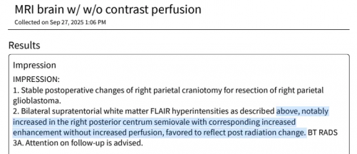
In the highlighted impression on the above radiology report, you’ll find the key phrase:
above, notably increased in the right posterior centrum semiovale with corresponding increased enhancement without increased perfusion, favored to reflect post radiation change. The centrum semiovale is deep white matter near the back of the brain, where communication pathways between brain regions run.
What?
The scan shows an area on the right side of the brain that appears slightly brighter on the MRI and exhibits some enhancement (from the contrast dye), but blood flow in this area hasn’t increased (that perfusion bit I described). That pattern typically indicates inflammation, scarring, or healing from prior radiation, rather than new tumor growth. Hence, the BT RADS 3A score that was a point of discussion in the prior post.
The overall guidance from the “neuro rad” (neuro radiologist who reads the scans and prepares the reports) to the ordering physician: Please keep a close eye on this with future scans.
Keep the Linear Accelerator Humming
The rub here is that (appropriately!), we have multiple specialists involved in the care of complex medical cases, for example, brain cancer! While the radiology report indicates post-treatment changes, things like functional status and clinical history matter, too, so my neuro oncologist offered their own interpretation and recommended a second opinion from my rad onc to determine if post-treatment changes are indeed validated or if we may be seeing disease progression.
1.21 gigawatts!
Yes, radiation therapy involves a linear accelerator, which is maintained by a medical physicist. That’s cool, right?! And if interested, 1.21 gigawatts is around the energy production of 2,600 Tesla Model 3s, or in a typical 1.5T MRI machine (1.5 T=tesla, as a unit of energy), you’d require 50,000 scans to use up a gigawatt of energy! Most hospital scanners are 1.5T-3T, with state-of-the-art scanners reaching up to 9T.
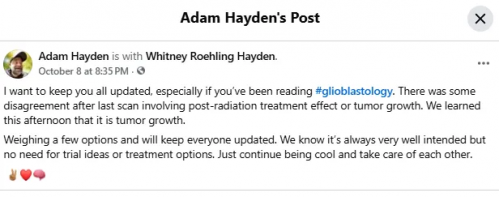
Anyway! My doc found the imaging to be more favorable for disease progression rather than treatment effect, and so, that’s the update we provided on Facebook and the reason we had the radiation consultation yesterday.
Courtesy of Adam Hayden/glioblastology.com
Cast in the Shadow of Illness
My favorite philosopher of health and illness—we all have one, right?!—is Havi Carel, and she wisely observed that in serious illness:
All social interactions are cast in the shadow of illness.
I suspect it resonates with many patients and care partners (caregivers) that the “sick role” includes becoming your own medical communicator to friends and family. Often, while navigating the earliest stages of diagnosis, a patient is thrust into the role of communicator to disseminate the info to their social network. This becomes complicated for several reasons!
- Patients are experts in experience, but we lack the technical expertise
- Serious illness is existentially distressing, and it becomes difficult to accept a diagnosis while helping others accept it, too
- Y’all want to spin up the best possible interpretation of results! We say “stable” and you say “remission”; we say “clean scan” and you say “no evidence of disease!” We say, “The chemo may be having a positive effect,” and you say, “Prayers answered.”
It’s not that we want to be negative, cynical, or resign ourselves to a defeatist attitude. It’s simply the case that over the past couple years, I’ve been on monthly chemo cycles, monthly MRI scan cycles, weekly abs, physical therapy, and so on. My daily meds treat seizures—I think most folks don’t understand that there is rarely a week that goes by that I am not involved in some sort of medical procedure, test, conversation, etc.
Cancer is nearly all-consuming.
And, as Prof. Carel says, it casts a shadow over all social interactions. I’m not sure I know any fellow patients who do not struggle with at least some of this medical communication to keep people appropriately updated, while delivering practical information, but not giving false hope or despair at the extremes.
And in my latest case, we’re discussing here: What is the right move? My doc indicated likely tumor growth and sent us to radiation.
Radiation, in fact, disagreed and cited the lack of hyperperfusion—the leaky blood—as strong evidence of post-treatment change and not disease progression, and so, we’re back where we were two weeks ago.
What Now?
Courtesy of Adam Hayden/glioblastology.com
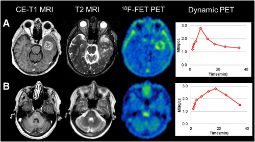
Comparison of multimodal brain imaging using contrast-enhanced T1-weighted MRI (CE-T1), T2-weighted MRI, and ^18F-FET PET with dynamic time–activity curves.
The View From FET PET
Here’s what comes next: another scan, this time a ^18F-FET PET, which looks at metabolism instead of anatomy. MRIs tell you what’s there — the shape of the scar, the swelling, the bright ring that could mean trouble. But after surgery, radiation, and chemo, the brain becomes tricky to read. Enhancement on a T1 or T2 can signal tumor… or just inflammation, blood vessels, or treatment effect.
See how this is exactly what we are now facing?
FET PET, an advanced imaging scan, examines behavior rather than structure. The scan is administered with a special compound, a “tracer” that active tumor cells soak up faster than normal tissue. What lights up here isn’t just tissue, it’s metabolism, the difference between what’s alive and what’s healing.
That distinction matters. FET PET helps tell disease from post-treatment change. In short, MRI shows what’s there; FET PET shows what’s active. And that’s what we’re trying to understand in my case.
The Next Month
Over the next month, we’ll continue to monitor my symptoms, paying close attention to functional decline—like my now required walker and wheelchair use, headaches, cognitive hiccups, etc. We are also waiting on the FET PET to be scheduled—this is still considered an “experimental agent,” and so, supplies are limited and timelines are tight.
Should we get scheduled, and should my health hold up with relative stability until that scan, we hope to discern post-treatment effects or disease progression. More of the waiting game that inevitably defines long-term survival. We simply do not have a large enough patient population in the associated clinical and radiographic longevity data to have standard treatment protocols.
Each day is a new data point — another line in the long experiment of living. Zoom out on that history, and you’ll see the real study underway: Whitney and me, raising our boys, showing up for family, preparing for what’s next, and keeping our community in the loop.
- <3 -a.
#AandWTumortakedown
This blog post was published by Glioblastology on October 11, 2025. It is republished with permission.


Comments
Comments